


- Oakstone Price: $995.
- Format: 39 Video Files (.mp4 format) + 2 PDF files.
- File Size: 12.3 GB
UCSF Abdominal and Thoracic Imaging 2019 (Videos+PDFs)
Stay Current with Key Advances
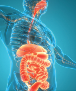
UCSF Abdominal and Thoracic Imaging is an extensive review of clinically relevant topics in chest, abdominal and ob-gyn imaging. This expertly designed course includes case-based lectures on topics like imaging of neuroendocrine tumors, female pelvis and early pregnancy, acute aortic syndrome, imaging of pleural disease, imaging mimics of pelvic pathology, etc. It will help you to:
- Identify common causes of acute pelvic pain in non-pregnant females
- Recognize the appearance of ectopic pregnancy
- Diagnose common and uncommon pancreatic masses
- Illustrate the proper management of a renal incidentaloma
- Apply updated guidelines for the diagnosis and management of lung nodules, lung cancer
- Differentiate pathologic conditions from benign incidentalomas and mimics in the lungs, heart and mediastinum
Expand Your Skills
Available online, UCSF Abdominal and Thoracic Imaging provides a maximum of 22.5 AMA PRA Category 1 Credits ™ and access to unbiased, evidence-based content and case-based reviews so you can expand your knowledge and incorporate the latest guidelines into your daily practice.
Accreditation
The University of California, San Francisco School of Medicine (UCSF) is accredited by the Accreditation Council for Continuing Medical Education (ACCME) to provide continuing medical education for physicians.
Designation
UCSF designates this enduring material for a maximum of 22.5 AMA PRA Category 1 Credits ™. Physicians should claim only the credit commensurate with the extent of their participation in the activity.
The total credits are inclusive of 12.25 in CT, 5.5 in MR, and 3.75 in Ultrasound.
Series Release: April 1, 2019
Series Expiration: March 31, 2022 (deadline to register for credit)
CME credit is obtained upon successful completion of an activity post-test and evaluation. CME Credit registration forms must be submitted prior to series expiration date.
Learning Objectives
Upon completion of this course, you will know how to:
- Diagnose common causes of acute pelvic pain in non-pregnant women
- Recognize the appearance of ectopic pregnancy
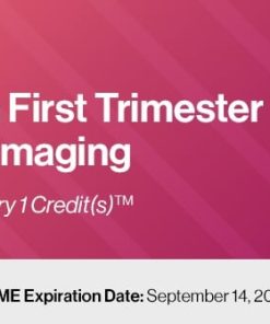
- Utilize ultrasound to identify findings suggestive and definitive of pregnancy failure in the first trimester
- Differentiate common solitary liver masses
- Describe the proper management of a renal incidentaloma
- Evaluate the small and large bowel in patients with abdominal pain
- Diagnose common and atypical pancreatic masses
- Apply updated guidelines for the diagnosis and management of lung nodules and lung cancer
- Recognize typical and atypical appearance of thoracic emergencies including pulmonary embolism and acute aortic syndromes
- Differentiate pathologic conditions from benign incidentalomas and mimics in the lungs, heart and mediastinum
- Apply standard terminology to improve description and classification of interstitial lung disease, small airways disease and bronchiectasis on HCRT
Intended Audience
This educational activity was designed for radiologists and other medical professionals who would benefit from a greater understanding of the interpretation of abdominal and thoracic images.
Topics/Speaker:
Female Pelvis and Pregnancy
- Imaging Mimics of Pelvic Pathology – Michael A. Ohliger, MD, PhD
- MRI of Gynecologic Malignancy – Michael A. Ohliger, MD, PhD
- Technical Tips & Tricks for MRI of the Female Pelvis – Michael A. Ohliger, MD, PhD
- Acute Pelvic Pain in the Non-pregnant Female – Lori M. Strachowski, MD
- Diagnosing Nonviable Pregnancy in the 1st Trimester – Lori M. Strachowski, MD
- Ectopic Pregnancy: Rings, Rules and Role of US – Lori M. Strachowski, MD
- O-RADS: An Introduction to the Lexicon and Risk Stratification – Lori M. Strachowski, MD
- US of the Uterus and Endometrium: Pearls to Perfect Your US Performance – Lori M. Strachowski, MD
Male Pelvis and Liver
- Hepatobiliary Agents and Their Role in Liver Imaging – Thomas A. Hope, MD
- Imaging of Biochemically Recurrent Prostate Cancer – Thomas A. Hope, MD
- LI-RADS Cases – Thomas A. Hope, MD
- MRI of Diffuse Liver Disease – Michael A. Ohliger, MD, PhD
- Prostate MRI T2 and Diffusion – Michael A. Ohliger, MD, PhD
- Rapid-body MR Imaging Techniques – Michael A. Ohliger, MD, PhD
- Sonography of the Scrotum: A Practical Approach – Lori M. Strachowski, MD
Gastrointestinal CT/MR
- Imaging of Neuroendocrine Tumors – Thomas A. Hope, MD
- Imaging of Rectal Cancer – Thomas A. Hope, MD
- CT of Peptic Ulcer Disease – Michael A. Ohliger, MD, PhD
- Acute Appendicitis – Emily M. Webb, MD
- CT of Colitis: Infection, Inflammation and Ischemia – Emily M. Webb, MD
- Cystic Pancreatic Masses: Imaging Update – Emily M. Webb, MD
- Everything You Need to Know about the Spleen in Less than an Hour – Emily M. Webb, MD
- Primary Retroperitoneal Masses: An Approach to Diagnosis – Emily M. Webb, MD
Abdominal and Thoracic
- Acute Aortic Syndrome – Travis S. Henry, MD
- Imaging of Pleural Disease – Travis S. Henry, MD
- Mediastinal Masses – Travis S. Henry, MD
- How to Handle Thoracic Incidentalomas – Kimberly G. Kallianos, MD
- Look at the Heart: Cardiac Findings on Non-gated CT – Kimberly G. Kallianos, MD
- Update on Pulmonary Embolism – Kimberly G. Kallianos, MD
- Acute Abdominal Pain – Emily M. Webb, MD
- Interesting Cases in the Abdomen and Pelvis – Emily M. Webb, MD
Thoracic and Pulmonary
- Bronchiectasis: An Imaging Approach – Travis S. Henry, MD
- Introduction to HRCT – Travis S. Henry, MD
- Lateral Chest Radiograph – Travis S. Henry, MD
- Lung Nodules: What Do I Ignore, and What Do I Further Explore? – Travis S. Henry, MD
- Small Airways Disease – Travis S. Henry, MD
- Challenging CXR Cases – Kimberly G. Kallianos, MD
- Confidently Diagnosing Lung Cancer – Kimberly G. Kallianos, MD
- Pulmonary Infections – Kimberly G. Kallianos, MD
Product Details
Only logged in customers who have purchased this product may leave a review.
Related Products

VIDEO MEDICAL
30 $

120 $

240 $

240 $


216 $

300 $
30 $
48 $

60 $

VIDEO MEDICAL
54 $

VIDEO MEDICAL
24 $
VIDEO MEDICAL
24 $
VIDEO MEDICAL
24 $
VIDEO MEDICAL
24 $
VIDEO MEDICAL
24 $
VIDEO MEDICAL
24 $
VIDEO MEDICAL
24 $
VIDEO MEDICAL
24 $
VIDEO MEDICAL
24 $
VIDEO MEDICAL
24 $
VIDEO MEDICAL
24 $

120 $
VIDEO MEDICAL
60 $
120 $

60 $

VIDEO MEDICAL
96 $


VIDEO MEDICAL
180 $
VIDEO MEDICAL
120 $

VIDEO MEDICAL
144 $

240 $
VIDEO MEDICAL
168 $

36 $

120 $
VIDEO MEDICAL
36 $
VIDEO MEDICAL
36 $
VIDEO MEDICAL
180 $
180 $

VIDEO MEDICAL
180 $

VIDEO MEDICAL
96 $

VIDEO MEDICAL
60 $
48 $

VIDEO MEDICAL
42 $
42 $


VIDEO MEDICAL
120 $

VIDEO MEDICAL
96 $


VIDEO MEDICAL
60 $

300 $
300 $
VIDEO MEDICAL
300 $
300 $
300 $
24 $
144 $

VIDEO MEDICAL
120 $
84 $

VIDEO MEDICAL
120 $

VIDEO MEDICAL
42 $

60 $

VIDEO MEDICAL
30 $


60 $

60 $

24 $

60 $

VIDEO MEDICAL
60 $
60 $

VIDEO MEDICAL
60 $

78 $

54 $

108 $

72 $

132 $
108 $

108 $

VIDEO MEDICAL
108 $
Reviews
There are no reviews yet.