

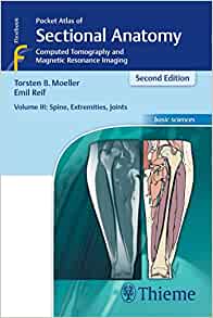
Atlas de Bolso de Anatomia Seccional – Tomografia Computadorizada e Ressonância Magnética – Volume III: Coluna Vertebral, Extremidades e Articulações: Volume 3, 1st edition
By Torsten B. Moeller
Known by all radiologists in the world as a reliable helper in light of its high quality illustrations and its practical features, the new edition of the Pocket Atlas of Sectional Anatomy incorporates the latest innovations in imaging technology, as well as hundreds of new illustrations. . It accurately and didactically describes the anatomical details seen in the images, presenting the corresponding schematic drawing in color, to facilitate the identification of anatomical structures. This is the perfect reference tool for radiologists and technicians working with cross-sectional imaging modalities. accompanied by explanatory drawings in color.
The Pocket Atlas of Sectional Anatomy is a must-have reference tool for radiologists and technicians working with cross-sectional imaging modalities. Authored by Torsten B. Moeller, this new edition incorporates the latest innovations in imaging technology, making it an indispensable resource in the field. With its high-quality illustrations and practical features, it is known and trusted by radiologists around the world.
The new edition of the Pocket Atlas of Sectional Anatomy features hundreds of new illustrations and accurately describes the anatomical details seen in the images. To facilitate the identification of anatomical structures, each description is accompanied by a corresponding schematic drawing in color. This visual approach makes it easier for radiologists and technicians to understand and interpret the images they encounter in their daily practice.
The book covers various imaging modalities, including computed tomography (CT), magnetic resonance imaging (MRI), and ultrasound. It provides comprehensive coverage of the anatomy of the head and neck, brain, spine, chest, abdomen, pelvis, and extremities. The detailed illustrations and clear descriptions help readers navigate through the complex anatomy of these regions.
One of the outstanding features of the Pocket Atlas of Sectional Anatomy is its practicality. The book is designed to be compact and portable, making it easy to carry and reference in any clinical setting. The spiral binding allows the book to lay flat, providing quick and easy access to the desired content. The organization of the book is intuitive, with each section focusing on a specific anatomical region.
In addition to its practical features, the Pocket Atlas of Sectional Anatomy is also a visually appealing resource. The illustrations are of high quality and showcase the anatomical structures in detail. The use of color enhances the clarity and understanding of the images, making it easier for readers to grasp complex anatomical concepts. The book also includes explanatory drawings that further clarify key anatomical structures.
Published by Thieme Revinter, the first edition of the Pocket Atlas of Sectional Anatomy was released on October 17, 2017. It is available in Portuguese language and is a digital eBook consisting of 480 pages. The ISBN-10 of the book is 856766196X and the ISBN-13 is 978-8567661964.
Why Order the Pocket Atlas of Sectional Anatomy?
The Pocket Atlas of Sectional Anatomy is an essential resource for radiologists and technicians working with cross-sectional imaging. Here are a few reasons why you should consider ordering this book:
1. Comprehensive Coverage:
The book covers the anatomy of various regions of the body, including the head and neck, brain, spine, chest, abdomen, pelvis, and extremities. It provides a comprehensive understanding of the anatomical structures encountered in cross-sectional imaging, allowing for accurate interpretation of images.
2. High-Quality Illustrations:
The book features high-quality illustrations that showcase the anatomical structures in detail. The use of color enhances the clarity and understanding of the images, making it easier to identify and interpret anatomical features.
3. Practical Design:
The Pocket Atlas of Sectional Anatomy is designed to be practical and user-friendly. Its compact size and spiral binding make it easy to carry and reference in any clinical setting. The intuitive organization of the book allows for quick and easy access to the desired content.
4. Updated with the Latest Innovations:
This new edition of the Pocket Atlas of Sectional Anatomy incorporates the latest innovations in imaging technology. It ensures that radiologists and technicians are up-to-date with the advancements in the field, enabling them to provide the best possible care to their patients.
5. Reliable and Trusted Resource:
The Pocket Atlas of Sectional Anatomy is known and trusted by radiologists worldwide. Its practical features, high-quality illustrations, and comprehensive coverage make it a reliable reference tool in the field. Whether you are a seasoned professional or a student, this book will be a valuable addition to your library.
Order Now:
If you are a radiologist or technician working with cross-sectional imaging modalities, the Pocket Atlas of Sectional Anatomy is a must-have resource. Its practical design, comprehensive coverage, and high-quality illustrations make it an invaluable tool in your daily practice. Stay updated with the latest innovations in imaging technology and enhance your understanding of anatomical structures with this trusted reference. Order your copy today and take your radiology practice to the next level!
Product Details
- Publisher : Thieme Revinter; 1st edition (17 October 2017)
- Language : Portuguese
- : 480 pages
- ISBN-10 : 856766196X
- ISBN-13 : 978-8567661964
Related Products
Anatomy Books PDF
Anatomie Und Physiologie Lehr- Und Übungsbuch Für Dummies (EPub)
Anatomy Books PDF
Anatomia Umana. Approccio Integrato Tra Struttura E Funzione (EPUB)
Anatomy Books PDF
Exploring Anatomy & Physiology In The Laboratory, 3rd Edition (Original PDF From Publisher)
Anatomy Books PDF
Exploring Anatomy & Physiology In The Laboratory, 3rd Edition (High Quality Image PDF)
Anatomy Books PDF
Lehrbuch Anatomie, 8th Edition (Original PDF From Publisher)
Anatomy Books PDF
Anatomy Books PDF
Neuroanatomy Text And Atlas, 5th Edition (Original PDF From Publisher)
Anatomy Books PDF
Sobotta Lerntabellen Anatomie Muskeln, Gelenke und Nerven, 4ed
Anatomy Books PDF
Anatomy Books PDF
Sobotta, Atlas der Anatomie Band 1: Allgemeine Anatomie und Bewegungsapparat, 25th ed
Anatomy Books PDF
Anatomy Books PDF

Anatomy Books PDF

Anatomy Books PDF
Anatomy Books PDF
Anatomy Books PDF
Anatomy Books PDF
Anatomy Books PDF
Anatomy Books PDF
Anatomy Books PDF
Textbook of Anatomy: Upper Limb and Thorax, Vol I, 4th edition
Anatomy Books PDF
Textbook of Anatomy: Abdomen and Lower Limb, Vol II, 4th edition
Anatomy Books PDF
Anatomy Books PDF
Anatomy Books PDF
Textbook of Anatomy-Head, Neck and Brain, Volume III, 4th edition
Anatomy Books PDF
Human Anatomy Laboratory Manual with Cat Dissections, 9th Edition

Anatomy Books PDF
Laboratory Manual for Hole’s Human Anatomy & Physiology, 16th Edition
Anatomy Books PDF

Anatomy Books PDF
Anatomy Books PDF
Anatomy Books PDF
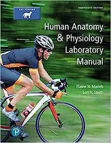
Human Anatomy & Physiology Laboratory Manual, Cat Version, 13th Edition
Anatomy Books PDF
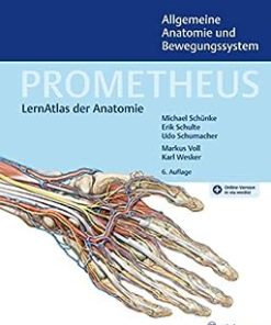
PROMETHEUS Allgemeine Anatomie und Bewegungssystem, 6th edition
Anatomy Books PDF
Anatomy Books PDF

Anatomy Books PDF
Anatomy Books PDF
Anatomy Books PDF
Memmler’s The Human Body in Health and Disease, Enhanced 14th Edition
Anatomy Books PDF
Anatomy Books PDF
Anatomy Books PDF

Anatomy Books PDF
Anatomy Books PDF
Anatomy Books PDF

Anatomy Books PDF
Anatomy Books PDF
Grant. Atlas de anatomía 15e (Spanish Edition) (High Quality Image PDF)
Anatomy Books PDF
Foundations of Anatomy and Physiology: A Workshop Manual with Laboratory Applications
Anatomy Books PDF

Anatomy Books PDF
Anatomy Books PDF
Anatomy Books PDF
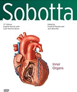
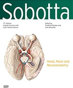
Sobotta Atlas of Anatomy, Vol. 3, 17th ed., English/Latin: Head, Neck and Neuroanatomy
Anatomy Books PDF
Anatomy Books PDF

Anatomy Books PDF

Anatomy Books PDF
Anatomy Books PDF
Anatomy Books PDF
Anatomy Books PDF
Anatomy Books PDF
Schaum’s Outline of Human Anatomy and Physiology, 4th Edition (Schaum’s Outline Series)
Anatomy Books PDF

Anatomy Books PDF
Anatomy Books PDF
Anatomy Books PDF
Anatomy Books PDF
Nicole Angemi’s Anatomy Book: A Catalog of Familiar, Rare, and Unusual Pathologies ()
Anatomy Books PDF
Anatomy Books PDF
BRS Gross Anatomy (Board Review Series), 10th Edition (EPUB3)
Anatomy Books PDF
Gray. Repaso de Anatomía: Preguntas y respuestas, 3 edición (Original PDF from Publisher)
Anatomy Books PDF
Anatomy: Regional, Surgical, and Applied (Original PDF from Publisher)
Anatomy Books PDF
Exercises for the Anatomy & Physiology Laboratory, 4th Edition (Original PDF from Publisher)
Anatomy Books PDF
Netter. Atlas de neurociencia, 4 Edición (Original PDF from Publisher)
Anatomy Books PDF
Moore’s Clinically Oriented Anatomy, 9th edition 2022 Original PDF

Anatomy Books PDF
Anatomy Books PDF
Selective Anatomy: Prep Manual for Undergraduates, 2nd edition, Vol 2 2020 Original PDF
Anatomy Books PDF
Selective Anatomy: Prep Manual for Undergraduates, 2nd edition, Vol 1 2020 Original PDF
Anatomy Books PDF
Clinical Kinesiology and Anatomy, Sixth Edition 2017 epub+converted pdf

Anatomy Books PDF

Anatomy Books PDF
Color Atlas of Human Anatomy: Vol. 2 Internal Organs, 8th edition 2022 Original PDF